Correct Presbyopia By Advanced FemtoLASIK PRESBYOND 10 minute(s) read
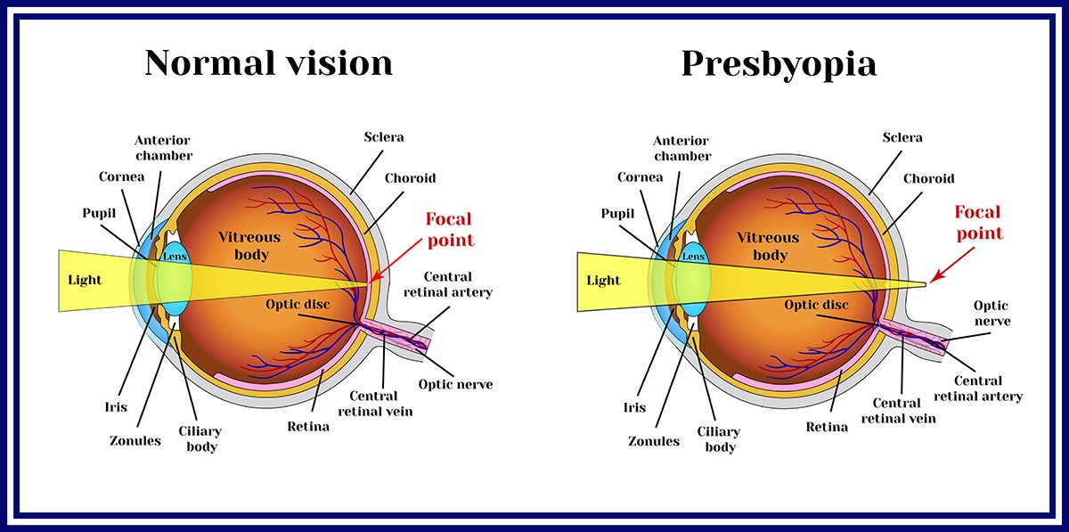
Presbyopia is defined as the gradual loss of eyes’ ability to focus on nearby objects. It is an age-related condition that usually becomes noticeable in an early to mid-40s. and appears to continue worsening as we age. Presbyopia is caused by a hardening of the lens of the eye, which occurs with aging. As the lens becomes less flexible, it can no longer change shape to focus on close-up images. As a result, these images appear out of focus, leading to a blurred vision at normal reading distance. The goal of presbyopia treatment is to compensate for the inability of the eyes to clearly focus on nearby objects. Instead of inconvenient methods, e.g. wearing corrective eyeglasses (spectacle lenses) or contact lenses, refractive surgery known as FemtoLASIK PRESBYOND is an effective treatment to reshape the shape of the cornea, resulting in an increased depth of focus.
Hyperopia
In a normally shaped eye, cornea and lens have a smooth curvature which refracts all incoming lights to generate a sharply focused image directly on the retina at the backsides of the eye. If a cornea or lens is not evenly curved, light rays cannot be refracted properly, causing a refractive error. Hyperopia occurs when an eyeball is shorter than normal or a cornea is curved too little (flattened cornea or decreased curvature). Therefore, the light is focused behind the retina, resulting in poor up closed or even poor distance vision. Hyperopia or farsightedness usually is present at birth and tends to run in the family. In addition, people with presbyopia are more likely to develop presbyopia earlier than others.
Presbyopia
Presbyopia is caused by a hardening of the lens of the eye. It is the gradual loss of near focusing ability that occurs with age. Most people begin to notice the effects of presbyopia after age 40. Presbyopia slowly develops due to the alterations in lens (decreased elasticity and increased hardness) and impaired ciliary muscle power of the eye, causing the eye to focus light behind rather than on the retina when looking at close objects. Presbyopia largely disrupts close-up works, such as reading, eating, using mobile phone or computer, driving, putting on make-up and sewing.
Symptoms Of Hyperopia Or Presbyopia
- Nearby objects appear blurry. Squinting is needed to see them clearly. If condition progresses, both distant and nearby objects become unclear.
- Eyestrain or headaches after reading or doing close-up work.
- Blurred night vision.
- Difficulty reading at normal reading distance.

Problems Caused By Hyperopia Or Presbyopia
The most common symptoms of presbyopia occur around age 40 for most people. However, presbyopia might arise earlier in some cases. Vision of seeing nearby objects becomes blurry, especially when the light is insufficient. Most people aged over 45 usually experience presbyopia which substantially interferes with their daily lives and activities. If it is left untreated, it can potentially lead to an impaired quality of life, low self-esteem and increased risk of accidents while driving or working with machines.
Visual Examinations
Hyperopia and presbyopia are diagnosed by a comprehensive eye examination, which includes:
- Ophthalmoscopy: An examination of the back part of the eye (fundus), which includes the retina, optic disc, choroid and blood vessels by using an ophthalmoscope.
- Tonometry: A diagnostic test that measures the pressure inside the eye, which is called intraocular pressure (IOP).
- Slit lamp: An instrument that consists of a high-intensity light source that can be focused to shine a thin sheet of light into the eye. It might be used in conjunction with other visual tests if needed.
- Corneal topography: A computer assisted diagnostic tool that creates a three-dimensional map of the surface curvature of the cornea. It is used to evaluate whether the curvature of the cornea is suitable for laser surgery.
- Phoropter or refractor: An instrument that contains different lenses used for refraction in order to measure an individual’s refractive error on each eye.
Once the results are obtained, an appropriate treatment plan for an individual patient can be designed based upon personal lifestyle and preference.
Treatments Of Hyperopia And Presbyopia
A wide range of treatment options include:
- Wearing corrective eyeglasses
- Wearing contact lenses
- Monovision
- FemtoLASIK PRESBYOND
- Conductive keratoplasty
- Scleral expansion surgery
- Intraocular lens implants (Implantable Collamer Lens or ICL)
- Wearing corrective eyeglasses
Principally, corrective eyeglasses for presbyopia consist of lenses with added magnifying power to compensate for an inability to see nearby objects.- For patients without previous vision problems, eyeglasses for near vision with the prescription of magnifying lenses should be worn while reading or looking at close objects. They should be removed when they are not needed.
- For patients with existing vision problems, such as nearsightedness, farsightedness or astigmatism, if corrective eyeglasses for farsightedness have been worn, but they are not improving the vision while looking at close objects, recommendations are:
- Wearing another corrective eyeglasses for near vision. However, it might be inconvenient since two eyeglasses need to be worn alternatively, causing headache and eye fatigue.
- Wearing corrective eyeglasses with bifocal lenses. These lenses have a visible horizontal line that separates distance prescription, above the line and reading prescription, below the line.
-
- In case of having nearsightedness, the previous prescribed corrective eyeglasses for long distance can also be used. If blurred vision happens when looking at nearby objects, removal of reading glasses might help since people with nearsightedness do not have vision issues while looking at nearby distance.
Even though wearing eyeglasses is a simple, safe way to correct vision problems, it can profoundly interfere with daily tasks and activities, e.g. golfing, running and swimming.
- In case of having nearsightedness, the previous prescribed corrective eyeglasses for long distance can also be used. If blurred vision happens when looking at nearby objects, removal of reading glasses might help since people with nearsightedness do not have vision issues while looking at nearby distance.
- Wearing contact lenses
- If contact lenses for nearsightedness, farsightedness or astigmatism have been regularly worn, corrective eyeglasses for farsightedness should be used to improve vision at close distance.
- Wearing monovision contact lenses. With monovision contact lenses, a contact lens for distance vision is worn in one eye (usually dominant eye) and a contact lens for close-up vision is worn in non-dominant eye.
- Monovision
With monovision, a contact lens needs to be worn on one eye to correct distance vision and another contact lens needs to be worn on other eye to correct near vision. The lens for distance vision is usually worn on the dominant eye. The patients normally use both eyes for distance and one eye for close-up work. This approach only works efficiently in 50% of the patients. However, patients may experience poor vision at night or in dimly lit environments. Some patients might have confusion or dizziness caused by a significant difference between two eyes.

- FemtoLASIK PRESBYOND
FemtoLASIK PRESBYOND, previously known as laser blended vision is an advanced laser eye surgery that aims to spontaneously correct a wide variety of vision problems, ranging from presbyopia, myopia (nearsightedness), astigmatism and hyperopia (farsightedness). Being independent from wearing corrective eyeglasses, FemtoLASIK PRESBYOND allows for an improved close-up, mid-range and distance vision in each eye, enabling the patients to freely continue their lives without any visual limitation.
FemtoLASIK is known as bladeless LASIK since all processes are conducted by using laser. FemtoLASIK PRESBYOND combines the simplicity and accuracy of corneal refractive surgery with the advantages of increased depth of field while retaining visual quality. Based on spherical aberrations, which naturally occur in the eye, it extends the scope of personalized ablation beyond the limits of conventional monovision laser methods. A customized ablation profile is designated for each eye to ensure optimized target refraction. Different optical zones can be selected to account for the patient’s pupil size.- In dominant eye: the curvature of the cornea needs to be slightly corrected in order to enhance the vision at mid-range and maintain the vision quality for far distance.
- In non-dominant eye: the curvature of the cornea needs to be extensively corrected in order to improve close-up and mid-range vision.
Key benefits of FemtoLASIK PRESBYOND
- Excellent visual acuity for near, far and intermediate distances without the use of corrective eyeglasses.
- Improved refractive outcomes with minimized side effects after surgery. The undesired effects of conventional monovision treatment, such as multiple images in one eye, seem to be entirely eliminated with FemtoLASIK PRESBYOND. It is an ideal approach that offers a physiologically optimized solution and represents a true binocular method for treating patients with presbyopia.
Patients who previously underwent LASIK for correcting myopia with poor distance vision, FemtoLASIK PRESBYOND can be further considered if the corneal thickness meets requirement for LASIK.
Nonetheless, the degeneration of ciliary muscle and lens involuntarily advances with age, therefore the effect of FemtoLASIK PRESBYOND on close-up vision can last approximately 7-8 years, depending on the strength of the ciliary muscles that control accommodation for viewing objects at varying distances.
- Conductive keratoplasty
Instead of using laser for treating presbyopia, conductive keratoplasty uses radiofrequency energy to contour the corneal by applying heat to tiny spots around the cornea. The heat causes the edge of the cornea composed of packed collagen fibrils to slightly shrink, increasing its curve or steepness and improving focusing ability. Despite that conductive keratoplasty is considered less-invasive procedure which can usually be completed within 2-3 minutes, the results are broadly variable and may not be long lasting. One to two years after receiving this treatment, the cornea often stretches and the effect of correcting farsightedness eventually disappears. - Scleral expansion
Scleral expansion surgery involves making small incisions in the eye and inserting bands to stretch the part of the sclera (the tough fibrous layer of the eyeball) that lies beneath the ciliary muscles that control accommodation. However, possible complications might include an eye irritation or inflammation. - Intraocular lens implantation
Intraocular lens implantation is a procedure which the synthetic lens is implanted in front of the natural lens to improve vision. This synthetic lens is so called implantable collamer lens or ICL.
Advances in technology have led to the development of numerous types of intraocular lenses, suitable for patients with different conditions, extending from hyperopia, myopia, astigmatism and presbyopia. ICL is a new intraocular lens that can be implanted into the eye without removing the natural lens. The lens is made from a material, called Collamer – a collagen co-polymer that contains a small amount of purified collagen. ICL is implanted through a microscopic incision and it does not alter the corneal curvature. Since the lens is soft, foldable and moist, it is considered highly safe and biocompatible, resulting in less chance of immunological reactions and inflammations. In addition, it minimizes halos around lights and glare at night. Moreover, the lens can be removed if required such as eyesight changes.
Although several types of lens implants are available for correcting presbyopia accompanied with other visual problems, an appropriate treatment plan for each patient present with different conditions must be made by an expert refractive surgeon.

Proper Eye Care
Despite the fact that hyperopia or presbyopia cannot be averted, proper eye care can greatly help maintain the vision acuity. These following instructions should be regularly followed:
- Using the 20-20-20 rule. The rule suggests that for every 20 minutes spent looking at a screen, a person should look at a distance 20 feet away for 20 seconds. This aims at reducing eye strain, blurred vision and dry eye caused by an overuse or looking at digital screens for too long.
- Having regular eye check-up. Annual eye examination is advised in order to evaluate visual clarity and detect if any abnormality arises.
- Wearing proper eyewear. Corrective eyeglasses must be worn as prescribed. New eyeglasses are needed if the vision changes over time.
- Being aware of any eye abnormality. If experiencing eye problems, e.g. blurred vision in one or both eyes, black spots or floaters and halos around lights, immediate medical attention must be sought.
- Protecting the eyes from UV light exposure. Too much exposure to UV light raises the risk of eye diseases. Wearing sunglasses that block UV radiations is crucial, especially for people who have long-term exposure to UV light or take certain drugs that induce photosensitivity
- Eating foods that benefit eye health and eyesight. Foods with nutrients essential for retinal protection and healthy eye are green leafy vegetables, e.g. spinach, kale and collard, carrot, sweet potato and cantaloupe.
- Smoking cessation. Studies have shown smoking increases the risk of eye diseases, including age-related macular degeneration, cataracts, glaucoma and dry eye syndrome. Only way to reduce the risk of developing these conditions is by refraining from smoking.
- Keeping underlying disease (s) under control. Since certain medical conditions, such as diabetes and hypertension can cause eye problems, thus these underlying diseases must be well controlled to avoid eye-related complications.
- Proper lighting for reading. To minimize eye strain, it is important to read books or do close-up tasks in proper lighting.
For people who seek for safe and effective treatments of myopia, hyperopia, stigmatism and presbyopia, FemtoLASIK PRESBYOND is an alternative choice that poses superior advantages for both close-up and distance vision. It is specifically recommended in patients with presbyopia in which their quality of life is significantly disrupted. To achieve the best possible outcome, refractive treatments require to be conducted by highly experienced and well-trained ophthalmologist.


